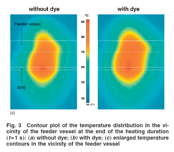
1. Anatomy of the eye and laser coagulation of choroidal feeder vessels. This figure gives the anatomic information of the eye. Specifically, the blood supply to the macular region of the retina is coming 100% from the underneath chriocapillary layer. When a patient develops age-related macula degeneration, abnormal growth of blood vessels may occur between the retina and the chriocapillary, therefore, disrupting mass transport between them and normal function of the retina. Using laser to directly close the abnormal growth of those small blood vessels may also permanently damage the retina. The approach proposed here is to aim the laser at the chroidal feeder vessel. The hypothesis is that the abnormal growth of blood vessels requires blood supply from the chriocapillary layer, laser coagulation of the feeder vessels will reduce the pressure variation in the chriocapillary layer, therefore, reducing the driving force to the abnormal growth. Clinical studies have demonstrated reduction or disappearing of the abnormal region after the closure of the feeder vessels by laser.
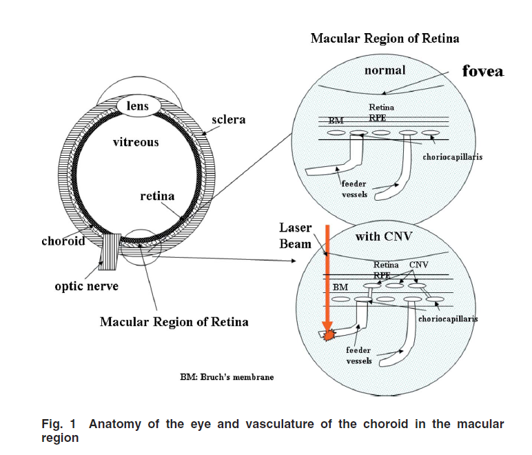
2. Temperature contours in the feeder vessel and its vicinity during laser irradiation have shown the temperature elevations in the targeted feeder vessel. The theoretical study has been used to design treatment protocols to achieve treatment efficacy while minimize collateral damage to surrounding healthy tissue.