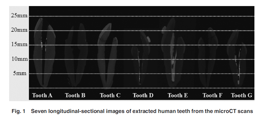
1. MicroCT images of extracted human teeth with a single root. The enamel and dentin are evident in the x-ray images. The bright region inside the root canal is due to root filling materials in dental procedures.

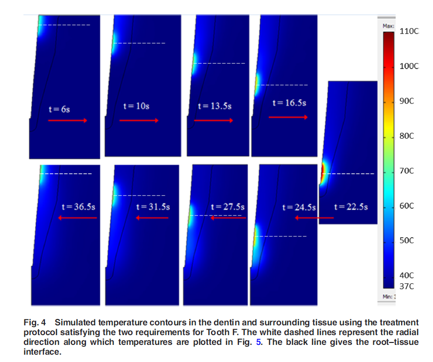
2. Temperature contours in tooth dentin during a designed laser heating protocol are presented in the following figure. The tooth root geometry is exported from the microCT images and imported into COMSOL software package. Note that the tooth root is embedded in a surrounding tissue. A laser catheter is moving inside the root canal from the cervical to apical regions and back to the cervical site. Significant temperature elelvations occur at the root canal surface, as well as in the deep dentin to eliminate bacteria there. The designed laser heating protocol allows minimal thermal damage to the interface between the root and its surrounding tissue.
